Considerations in Drug Design

Drug design is a sophisticated pharmaceutical discipline with a rich history of achievements. Since the late 19th century, when Emil Fisher proposed the concept of drug-receptor interaction as a key and lock mechanism, significant progress has been made in the field. Over time, drug design has evolved into a well-structured science with a solid theoretical foundation and practical applications. Today, it stands as the leading approach to drug discovery, leveraging advancements in science and technology to develop effective, specific, non-toxic, safe, and well-tolerated medications.

The cornerstone of any successful project lies in the meticulous design and thorough planning that take into account all potential factors that could impact its outcome. This principle is especially crucial in the realm of intelligent drug design, where the key to successful drug discovery and development lies in identifying molecules with beneficial characteristics. However, the process is highly complex, time-consuming, and resource-intensive, necessitating multi-disciplinary expertise and innovative approaches influenced by numerous critical factors. Without comprehensive drug design, there is a risk of failing to obtain FDA approval after investing substantial time and resources. Medicinal chemists play a pivotal role in conscientiously designing drug candidates, drawing upon their specialized knowledge and expertise to navigate the drug discovery process.

Several steps are involved in drug development, including:

  • Discovery and development: Research for a new drug begins in the laboratory
  • Preclinical research: Drugs undergo laboratory and animal testing to evaluate safety
  • Clinical research: Drugs are tested on people to ensure safety and effectiveness
  • FDA review: FDA review teams thoroughly assess all submitted data to determine approval
  • FDA post-market safety monitoring: FDA monitors drug and device safety after products are available for public use

The process of drug discovery begins with the identification of a hit molecule, which demonstrates a desired activity in a screening assay. Subsequently, the structure of this molecule is refined to enhance affinity and selectivity, minimize toxicity, improve solubility in water and lipids, enhance general ADME properties, and transform the hit molecule into a lead molecule. Further optimization of the lead molecule culminates in the development of a drug candidate for preclinical trials.

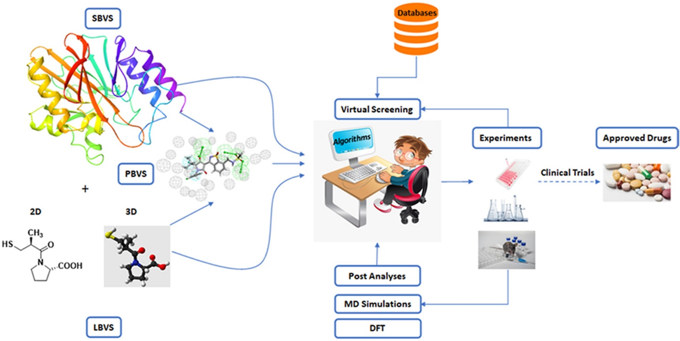
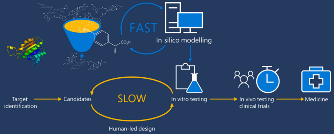
Medicinal chemists start drug design in the Discovery and Development phase. While advancements have been made in the modernization of the drug discovery process, most of the key factors for drug design have remained consistent. The integration of artificial intelligence (AI) and deep learning (DL) has recently accelerated the drug design process, improving efficiency and effectiveness. There are two main types of drug design: ligand-based drug design (LBDD) and structure-based drug design (SBDD). SBDD can be further categorized into de novo design and virtual screening, while LBDD can be categorized into QSAR, scaffold hopping, pseudo receptors, and pharmacophore modelling as illustrated in Figure 1 [1].

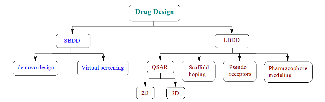
Figure 1: Types of drug design

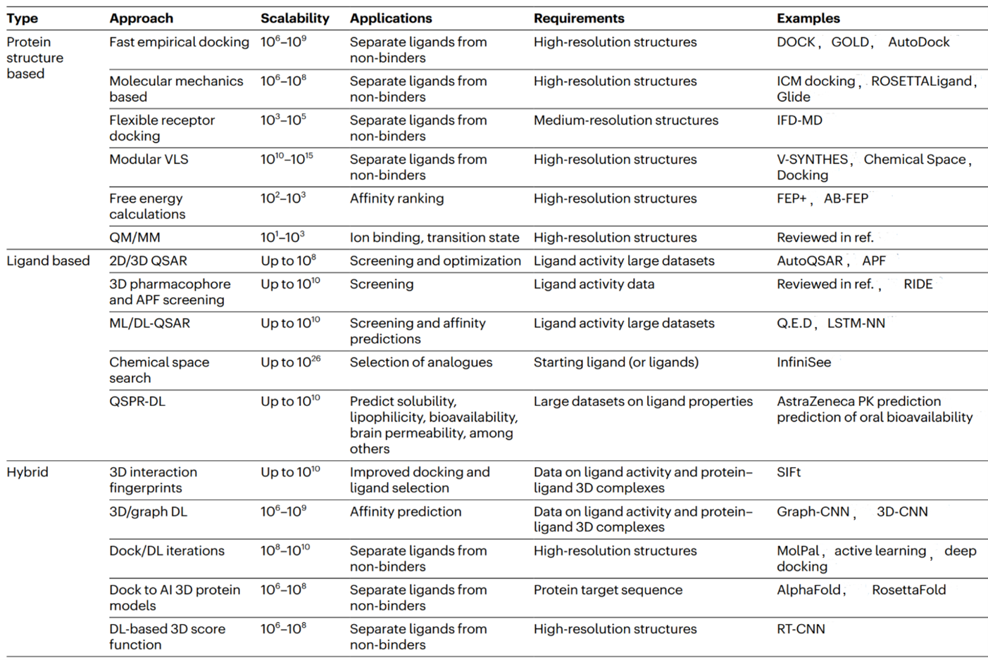
The elucidation of protein structures is achieved through various techniques such as X-ray crystallography, NMR spectroscopy, and cryogenic electron microscopy, with data being stored in the Protein Data Bank (PDB), which currently holds over 180 thousand structures. While the majority of these structures are single proteins in their apo-form, a subset includes complexes with ligands, providing valuable information on protein-binding sites. A significant advancement in modern drug design is the utilization of in silico modeling technologies for virtual screening, compound design, energy calculations, SAR and QSAR analysis, ADME modeling, and drug-target interaction modeling. To leverage these advanced technologies, molecular structures must be numerically encoded to enable analysis, search, visualization, and comparison. Encoding can be done using binary strings, smiles strings, 2D graphs, and 3D structures. The incorporation of 3D molecular modeling and visualization is recognized as a significant achievement in drug design technologies.

Computer-aided drug design (CADD) is widely recognized as a powerful tool in the drug discovery process. Techniques such as structure-based and ligand-based drug design through CADD provide crucial insights for molecular docking, molecular dynamics, and ADMET. Molecular docking is a computational method used to determine the precise binding pose of a protein-ligand complex and assess the strength of the interaction. AI and its subsets, Machine Learning (ML) and Deep Learning (DL), play a significant role in modern drug design by using vast datasets found in academic journals, patents, books, dissertations, reports, conferences, and clinical trials to generate novel and promising drug molecules.

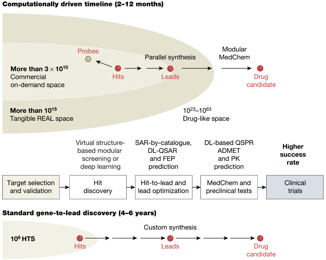
The concept of sequence-to-drug represents an innovative approach in computational drug design that leverages protein sequence data through end-to-end differentiable learning. This cutting-edge methodology utilizes Transformer models such as CPI2.0 and AlphaFold as foundational tools, showcasing their ability to generalize across proteins and compounds. It is crucial to interpret the binding knowledge obtained from Transformer models to aid in the discovery of new hits for challenging drug targets and to identify new targets for existing drugs through reverse application. Unlike traditional drug design projects that involve a complex, human-engineered pipeline with independently optimized steps, this new concept streamlines the entire learning process in a self-consistent and data-efficient manner, potentially minimizing error accumulation in complex pipelines (Figure 2) [2].

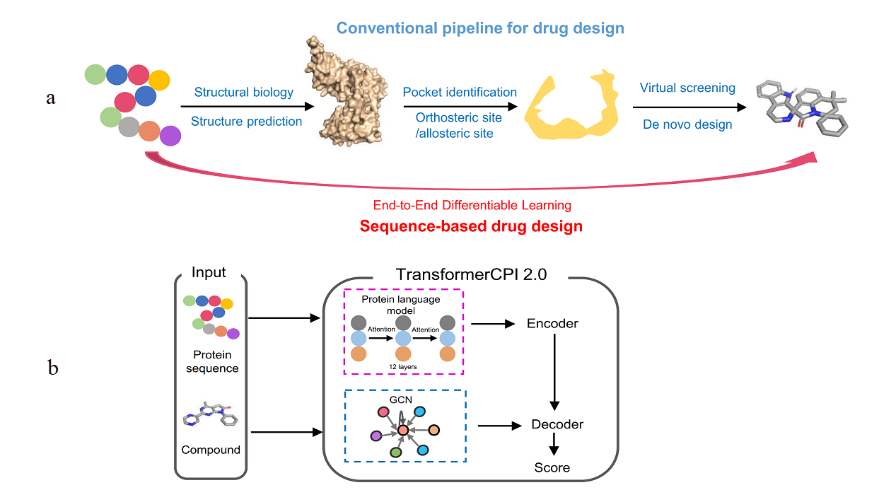
Figure 2:  a) The conventional pipeline for target-based drug design and the sequence-to-drug concept;  b) The computational pipeline of TransformerCPI2.0

The utilization of fragment linking as a valuable tool for the rational design of drug leads has been widely recognized in both academic and pharmaceutical settings. This approach is commonly employed to develop small molecule protein inhibitors and potential drug candidates. The initial stage of the fragment-based drug discovery process involves identifying fragments that exhibit weak binding affinity to the target protein, typically in the micromolar to millimolar range. Fragments are characterized as low molecular weight (<300 g/mol) and highly soluble organic molecules. Subsequently, these fragments are connected by a linker and optimized to create a single molecule with enhanced potency and improved drug-like properties. Two illustrative examples are provided in Figure 3 [3].

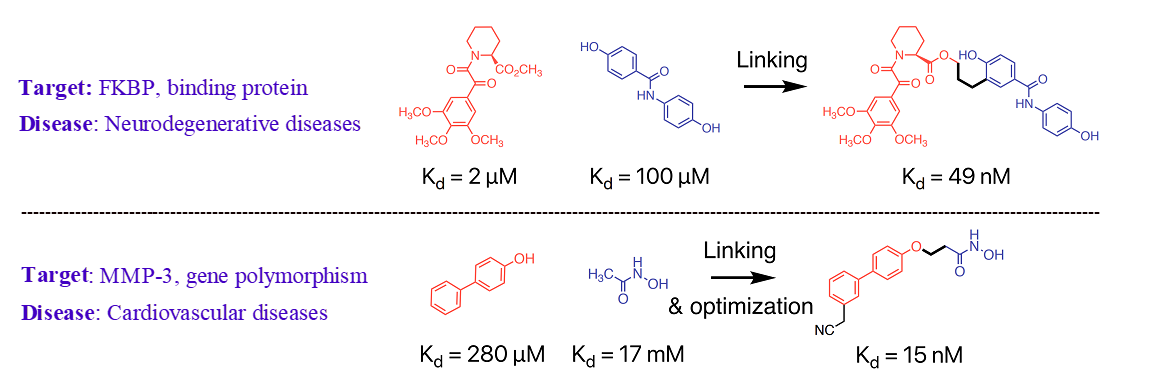
Figure 3: Fragment linking and optimization

In the field of drug discovery and development, Lipinski’s Rule of Five is a crucial consideration when designing small molecule drug candidates. This rule assists in predicting whether a biologically active molecule will have the necessary chemical and physical properties for oral bioavailability. The Rule of Five is based on specific physicochemical properties that impact pharmacokinetic drug properties such as absorption, distribution, metabolism, and excretion. According to Lipinski’s Rule of Five, orally active drugs generally should have no more than 5 hydrogen bond donors (HBDs), no more than 10 hydrogen bond acceptors (HBAs), a molecular mass less than 500 Da, and a partition coefficient (Clog P) not greater than 5. In drug design, molecules of interest generally exhibit a lower number of HBDs compared to HBAs. However, published data analyses for drug-like compounds do not definitively support the notion that HBDs pose greater challenges than HBAs in terms of ADME [4]

In the field of medicinal chemistry, bioisosteres are chemical substituents or groups that exhibit similar physical or chemical properties, resulting in comparable biological effects within the same compound. Utilizing bioisosteres in drug discovery offers a strategic and effective method to enhance molecule properties, such as increasing potency, addressing pharmacokinetic challenges, minimizing off-target effects, and optimizing physicochemical characteristics. The utilization of bioisosteres in refining bioactive compounds is a widely recognized and continually evolving approach that fosters innovation. For example, an illustration of bioisosteres related to carboxylic acid can be seen in Figure 4 [5].

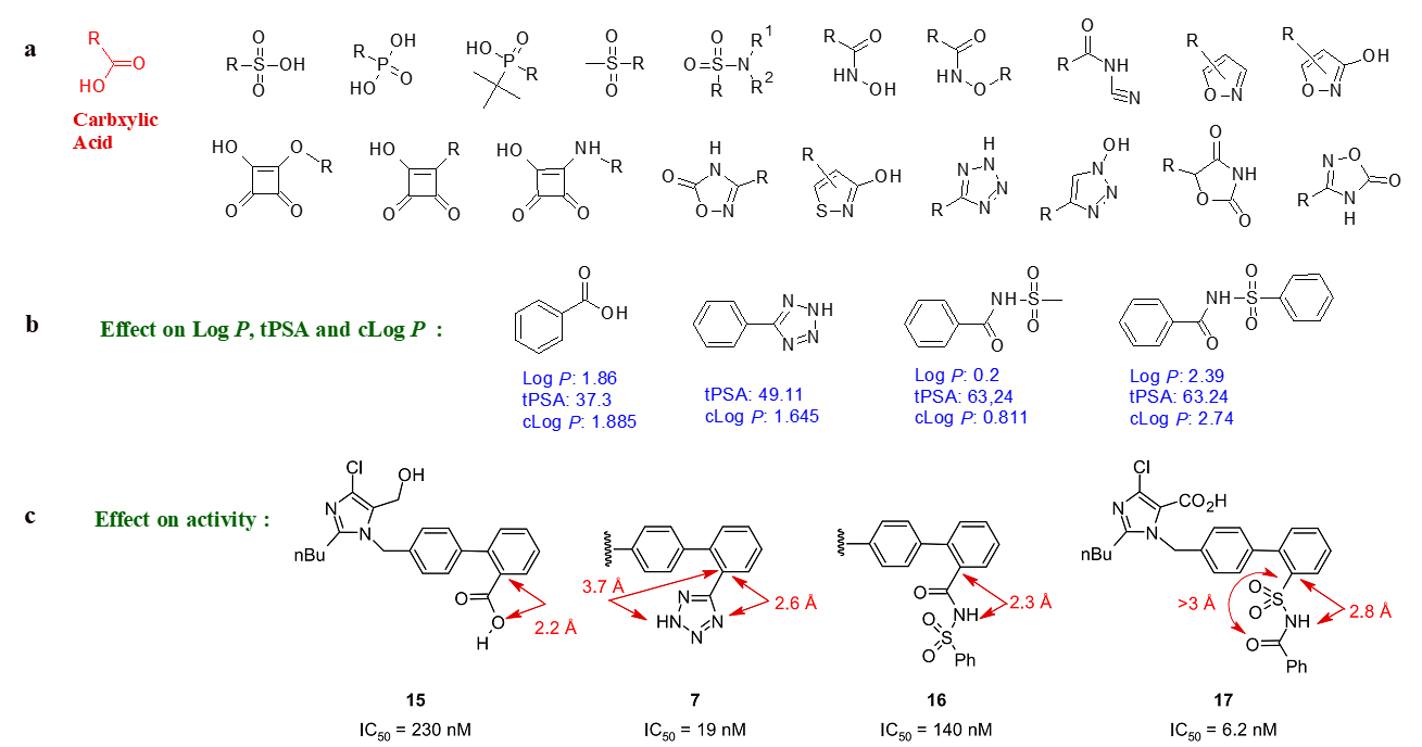
Figure 4: a) Examples of carboxylic acid bioisosteres;  b) Bioisosteres: effect on properties;  c) Bioisosteres: effect on bond length and activity

The process of drug design is a powerful tool within the pharmaceutical industry for discovering new drug leads against important targets. However, designing and synthesizing organic molecules with specific pharmaceutical applications presents a significant challenge, as it requires consideration of various factors such as selectivity, potency, pharmacokinetic properties, and scalability. It is worth noting that the majority of drug design approaches heavily rely on reported biological activity and properties data, which are often limited in quality and reliability. The drug design process is predominantly theoretical, which may result in discrepancies between expected and actual outcomes. Furthermore, while designing molecules may seem straightforward, their laboratory preparation can be quite challenging.

Innovative and cost-effective strategies are necessary for advancing drug discovery, including the utilization of artificial intelligence to efficiently analyze vast datasets for target identification and ligand selection. The ultimate objective of drug development is to create personalized drugs that are safe, effective, and rapidly developed. While this goal may seem ambitious, it is certainly achievable in the near future.

References:
[1] Layla Abdel-Ilah, Elma Veljović, et al; IJERT 2017, 6, 582-587
[2] Lifan Chen, Zisheng Fan Jie Chang, et al; Nat. Commun. 2023, 14, 4217
[3] Alexandre Bancet, Claire Raingeval, et al; J. Med. Chem. 2020, 63, 11420-1435
[4] Peter W. Kenny; J. Med. Chem. 2022, 65, 14261-14275
[5] Nicholas A. Meanwell; J. Agric. Food Chem. 2023, 71, 18087-18122

Computational Approaches in Drug Discovery

The process of drug discovery is an intricate, expensive, and time-consuming endeavor with a notably low success rate due to the complex nature of biological systems involving diverse heterogeneous elements that interact with each other, forming subsystems at different organizational levels. Consequently, extensive research has been conducted on molecular-level interactions within the context of disease mechanisms and drug discovery, resulting in the development of computational approaches. As a result, numerous databases provide readily available information on the relationships between drugs, diseases, proteins, and related concepts. Computational applications and tools have been widely adopted since the early 1980s to facilitate and expedite the discovery of drugs, giving rise to the field of computer-aided drug design (CADD). Over time, advancements in theoretical chemistry, physics, computer science, and statistics, coupled with the exponential growth in computational power, have greatly contributed to the field’s progress. Additionally, the evolution of data science has facilitated the transformation of extensive knowledge, enabling the emergence of bioinformatics and cheminformatics.

Computational methods are primarily employed in the early stages of drug discovery, where they play a crucial role in understanding disease biology, prioritizing drug targets, and optimizing chemical entities for therapeutic intervention. The main objectives of in silico approaches in drug discovery are to generate improved compounds with desirable in vitro and in vivo properties. Additionally, computational analysis aids in decision-making and guides experimental programs, reducing the number of candidate compounds that need to be evaluated experimentally. Presently, computational approaches have become indispensable tools at all stages of the drug discovery and development process. There are various computational methodologies available to assist researchers in identifying and investigating potential new drug candidates. Notably, structure-based drug design (SBDD) and ligand-based drug design (LBDD) are predominantly employed for drug design through CADD. Table 1 [1] provides a comprehensive overview of the primary computational approaches used to screen protein targets for potential ligands.

Table 1:  Major types of virtual screening algorithms

With the prolonged development period, increased cost, and limited probability of success, the rapid and effective formulation and synthesis of novel drug molecules pose significant challenges. The drug discovery process typically entails a design-make-test-analyze (DMTA) cycle lasting 10 to 15 years and incurring an average cost exceeding billion dollars to bring a new drug to the market. To mitigate these challenges and optimize time, resources, and risk, the incorporation of CADD methodology has become widespread. Research indicates that the utilization of CADD techniques can result in a 50% reduction in drug research and development costs. Additionally, the implementation of computationally driven drug discovery significantly shortens the identification time for potential drug candidates compared to traditional synthesis-driven discovery, as depicted in Figure 1 [1].

Figure 1. Computationally driven drug discovery. Schematic comparison of the standard HTS plus custom synthesis-driven discovery pipeline versus the computationally driven pipeline.

The field of drug discovery relies on several essential computational approaches, such as SBDD, LBDD, virtual screening, molecular docking, pharmacophore, quantitative structure-activity relationship (QSAR), and absorption, distribution, metabolism, excretion, and toxicity (ADMET).

  • SBDD involves the calculation of the interaction or bio-affinity between tested compounds and proteins with known three-dimensional (3D) structure. This enables the design of therapeutic molecules with improved interactions with target proteins.
  • LBDD, on the other hand, focuses on target proteins with unknown 3D structure but known ligands that bind to the intended target location. Through docking, these ligands can be used to create a pharmacophore model or molecule with the necessary structural characteristics to bind to the target active site. LBDD considers substances with similar structural similarities to have comparable biological actions and interactions with the target protein.
  • Virtual screening has become a convenient tool for identifying bioactive compounds. It utilizes information about the protein target or known active ligands. There are two types of virtual screening approaches: structure-based virtual screening (SBVS) and ligand-based virtual screening (LBVS).
  • Molecular docking (Figure 2) [2] is an in-silico approach used to predict the location of small molecules or ligands within the active region of their target protein. It accurately estimates the most favorable binding modes and bio-affinities of ligands with their receptors. Prediction of binding posture, bio-affinity, and virtual screening are the three primary goals of molecular docking, which are interconnected. The search and scoring algorithms used in molecular docking are fundamental tools for generating and evaluating ligand conformations.

Figure 2. Molecular docking: (a) Small-molecule ligand binding, (b) peptide binding and (c) protein–protein interactions

  • Pharmacophore refers to a schematic representation of bioactive functional groups and their interatomic distances.
  • QSAR is a technique employed when structural-based approaches are not applicable due to a lack of knowledge about the target macromolecule structure. It provides information on the relationship between chemical structure and biological activity. The main advantage of QSAR is its ability to identify characteristics of new chemical compounds without the need for production and testing.
  • ADMET evaluation of leads in the early stages of drug screening is necessary to address high attrition rates caused by poor pharmacokinetic profiles. Virtual screening can be utilized to filter hits and eliminate compounds with undesirable properties before comprehensive experimental testing. In silico ADMET filters, such as QSAR, use chemical or molecular descriptors to predict drug-like characteristics of compounds. ChemBioServer offers features such as displaying and graphing molecular characteristics, filtering compounds based on chemical quality, steric conflicts, and toxicity, searching for substructures, clustering compounds, and proposing a representative for each group.

The advancement of generative chemical spaces can be further facilitated by innovative computational methodologies in synthetic chemistry. For instance, the utilization of deep learning-based retrosynthetic analysis enables the prediction of novel iterative reaction sequences, synthetic routes, and feasibility. The discovery process of over 70 commercialized drugs included the use of computational techniques, as outlined in Table 2 [3] for example. It is important to note that in most cases, the initial drug lead (rather than the final commercial drug) was discovered with the aid of computer-aided drug design (CADD) techniques.

Table 2: Commercial drugs that made use of CADD during the discovery process

Drug Biological action Computational contribution to drug discovery Approval
Captopril Angiotensin converting enzyme (ACE) LBDD (Ligand-based drug design) and Structure-activity relationship (SAR) and SBDD (Structure-based drug design) 1981
Norfloxacin Topoisomerase II, IV SBVS, LBDD and QSAR Modelling 1986
Imatinib Tyrosine kinase SBDD (chemical libraries were screened for inhibitors against Bcr-Abl tyrosine kinase) 1990
Epalrestat Aldose Reductase MD and SBVS 1992
Cladribine Adenosine deaminase SBDD (VS and Docking) 1993
Saquinavir HIV-1 protease SBDD (Transition-state mimetic concept) 1995
Indinavir HIV-1 protease SBDD (Transition-state mimetic concept guided by molecular modelling and X-ray crystal structure) 1996
Zanamivir Influenza Neuraminidase  SBDD (Computer-assisted modelling of the active site) 1999
Lopinavir HIV-1 protease SBDD (Transition-state mimetic concept) [3D modelling and docking. Energy minimization using DISCOVER CVFF force field] 2000
Eptifibatide Glycoprotein IIb/IIIa Peptide-based (barbourin) design 2001
Valsartan Angiotensin II receptor Superimposition of energy-minimized conformation and QSA 2002
Enfuvirtide HIV-1 protease Homology Modelling 2003
Erlotinib EGFR kinase SBVS 2005
Ambrisentan Endothelin-A receptor SBDD (Docking), FBDD and Virtual Screening 2007
Raltegravir HIV-1 integrase Combining MD with flexible-ligand docking 2007
Tomudex Thymidylate synthase SBDD 2009
Crizotinib ALK and ROS1 SBDD and SAR 2011
Rivaroxaban Factor Xa HTS, SBDD and Virtual SAR 2011
Dolutegravir HIV-1 Integrase PBDD (two-metal binding pharmacophore structural based design) 2013
Saroglitazar PPAR Combined virtual screening of 3D databases, SBDD and Pharmacophore Modelling 2013
Grazoprevir NS3/4 A protease Molecular Modelling and Docking-derived approach 2016
Rucaparib Poly (ADP-ribose) polymerase (PARP-1) Ligand-based molecular modelling 2016
Acalabrutinib Bruton’s tyrosine kinase SAR, SBDD and Docking 2017
Betrixaban Serine protease Factor Xa (fXa) Molecular Docking 2017
Brigatinib ALK Docking and Homology Modelling 2017
Copanlisib HCl Phosphoinositide 3-kinase (PI3K) SBDD (Xray crystallography and Docking) and LBDD (based on lead scaffold) 2017
Vaborbactam β-Lactamase Docking and MD 2017
Duvelisib PI3K Kinase SBDD (Molecular docking, virtual screening) and LBDD (lead optimization and SAR) 2018
Apalutamide Androgen receptor inhibitor SBDD and SAR 2018
Dacomitinib Oral kinase Combined FBDD and SBDD 2018
Talazoparib Tosylate Poly (ADP-ribose) polymerase-PARP SBDD, SAR and Lead Optimization 2018
Darolutamide Androgen receptor SBDD (Docking and MD) 2019
Erdafitinib FGFR tyrosine Combined FBDD and SBDD 2019
Fedratinib HCl Tyrosine kinase SBDD (Virtual screening and Molecular Docking) 2019
Selinexor Nuclear export SBDD (consensus induced fit docking 2019
Zanubrutinib Bruton’s tyrosine kinase inhibitor Combined FBDD and SBDD 2019

The profound impact of computer-aided drug design (CADD) on pre-clinical drug development is evident. Furthermore, the application of CADD is continuously expanding alongside advancements in virtual screening and molecular docking. The integration of molecular dynamics simulations and experimental evaluation has the potential to enhance the reliability of refining computational models for identifying promising prospective hits. It is important to highlight that computational approaches continue to be a crucial resource in pre-clinical drug development, with a proven track record in facilitating the development of new drugs and repurposing existing ones.

Despite the numerous advancements and applications in drug discovery and development utilizing computational approaches, there are still several unresolved obstacles and challenges. These include the lack of synergistic computational models, quality datasets, standardization, accurate scoring functions, issues with multi-domain proteins, and assessment of multi-drug effects. Additionally, improvements in specific areas are necessary, such as enhancing the efficiency of virtual screening, increasing the quantity and quality of online computational resources, further advancing computational chemogenomics, designing drugs for multiple molecular targets, enhancing predictive toxicity models and side effects, and  collaborating with other disciplines to optimize the search for bioactive compounds for treating and preventing diseases.

References

[1] Anastasiia V. Sadybekov and Vsevolod Katritch; Nature 2023, 616, 673-685.
[2] Jinan Wang, et al; QRB Discovery 2022, 3: e13, 1–12.
[3] Victor T. Sabe, Thandokuhle Ntombela, et al;  Eur. J. Med. Chem. 2021, 224, 113705.

Late Stage Functionalization in Drug Discovery

A minor functional modification to the structure of a drug candidate can have a profound impact on its biological profile. However, the optimization process in the final stage of drug development is often costly, complex, and time-consuming, especially when a complete de novo synthesis is required for structure activity relationship (SAR) based or artificial intelligence (AI)-aided target molecules. To address this issue, the recent concept “Late Stage Functionalization” (LSF) offers a potential solution. LSFs are highly versatile methodologies that effectively perform desired chemoselective transformations on intricate molecules to deliver numerous diverse products in a single reaction.  LSF, especially when applied to the diversification of C-H bonds, presents a promising opportunity to explore new chemical space, particularly with sp3-carbon atoms. For late-stage diversification to become a valuable tool in drug discovery, advancements in high-throughput technologies and predictive techniques are necessary to accelerate the biological testing of the leads generated through this approach.

Drug discovery and development is a laborious, exceedingly costly, and highly precarious undertaking. The clinical progression of a drug candidate, spanning from the initiation of a clinical trial to obtaining marketing approval, demonstrates a discouragingly low rate of success (10%-20%) and necessitates a substantial investment, thereby demanding significant incentives for pioneering advancements in chemical synthesis to enhance production efficiency. Recently, the LSF reaction methodology has advanced to the point where it has started to have an impact in the field of medicinal chemistry. It can now be effectively utilized for reactions that require mild conditions and tolerance of various sensitive functional groups. In the past decade, there has been a significant increase in publications on various late-stage transformations, as evidenced by a comprehensive literature search conducted in Scopus, Web of Science, and SciFinder, as shown in Figure 1 [1]. The analysis of this literature search indicates that the present LSF toolbox encompasses numerous chemical and enzymatic methods, including fluorination, amination, hydroxylation, and methylation.

Figure 1: Increasing use of LSF.

LSF is a valuable tool for accessing derivatives that would otherwise be challenging or time-consuming to produce. Its utility extends to various applications, including SAR of pharmaceutical candidates and the creation of molecules with unstable isotopes that would not survive long enough for traditional synthesis methods. While LSF cannot replace de novo synthesis in all cases, it offers a means to access molecules that would have otherwise remained undiscovered. The ability to selectively diversify structurally complex molecules at a late stage holds significant potential for drug discovery. The desired features and useful tools of LSF have been demonstrated in Figure 2 [2]. In recent years, medicinal chemists have incorporated LSF strategies into their drug discovery programs, resulting in efficient access to diverse libraries for exploring structure-activity relationships and improving physicochemical and pharmacokinetic properties. Notably, substantial advancements have been made in providing reaction conditions for functionalizing sp2 or sp3 C-H bonds without the need for a synthetic handles.

Figure 2: Desired LSF transformations and required features

The functionalization of C-H bonds offers significant opportunities for more efficient exploration of chemical space compared to traditional synthetic strategies, eliminating the need for extensive redesign of synthetic routes. Specifically, C-H bond late-stage functionalization enables the direct and selective conversion of prevalent C-H bonds into various C-X bonds (such as C-C, C-N, C-O, C-halogen) in complex molecular structures, without the requirement of pre-installing a functional group. This approach presents a straightforward and cost-effective method to accelerate the optimization of ADME profiles, physical properties, and the identification of structure-activity relationships, ultimately enhancing on-target potency and reducing off-target effects. Notably, the conversion of a C-H bond to C-CH3, known as the “Magic Methyl” effect (Figure 3) [3], exemplifies the profound pharmacological effects achievable. The ability to enable late-stage C-H functionalization in bioactive molecules holds significant value for drug discovery and development.

Figure 3: Magic Methyl Effect

Despite the significant progress made, strategies for LSF are still in the early stages of development. There are several challenges that need to be addressed in order to expand the range of methodologies applicable to pharmaceuticals. The functionalization of a drug candidate through C–H bonds may be limited due to selectivity issues that arise in complex molecules, or the lack of existing methodologies for the desired C–H functionalization. Most drug-like molecules have multiple reactive sites and their own complexities, resulting in a distinct reactivity pattern for each compound. Consequently, it is challenging to provide a comprehensive method that can overcome all the practical difficulties associated with LSF approaches. Currently, LSF of complex molecules is an immature approach with limited scope and success rates, often accompanied by inadequate control over chemoselectivity and/or regioselectivity.

References

[1] David F. Nippa, Remo Hohler, et al; Chimia 2022, 76, 258–260
[2] Lucas Guillemard, Nikolaos Kaplaneris, et al; Nat. Rev. Chem. 2021, 5, 522-545
[3] Edna Mao and David W.C. MacMillan; J. Am. Chem. Soc. 2023, 145, 2787−2793

FDA Approved Drugs Using Lipid Nanoparticles Mediated Payload Delivery

Nanomedicine, the field that encompasses the convergence of nanotechnology, pharmaceutical, and biomedical sciences, has experienced rapid advancements in recent years. Particularly notable is the development of new nanoformulations for therapeutic purposes, imaging agents, and theragnostic applications. One area of great promise within nanomedicine is the use of lipid-based nanoparticles for targeted delivery of nucleic acids to disease-causing active sites. These offer solutions to common challenges in drug delivery, such as low water solubility and poor bioavailability. Furthermore, lipid-based nanoparticles have the ability to overcome various physiological obstacles, enabling enhanced distribution to the desired sites. Recently, lipid nanoparticles (LNPs) have proven successful in efficiently delivering cytotoxic chemotherapy agents, antibiotics, and nucleic acid therapeutics. The significance of this field has been recognized by awarding the Nobel Prize of 2023 in Physiology or Medicine to Katalin Karikó and Drew Weissman for their discoveries enabling the creation of a new type of messenger RNA (mRNA) vaccine using LNPs as delivery vehicles. The mechanism of action of LNP-mRNA mediated vaccination is depicted in Figure 1 [1].

Figure 1. Mechanism of action of LNP-mRNA mediated vaccination.

RNA therapeutics show great potential in various medical applications, including virus vaccines, cancer immunotherapy, and gene editing. Various drug delivery vehicles, including LNPs, have been developed to address the instability of RNA. LNPs have been regarded as the most efficient and suitable delivery system for nucleic acids, such as small interfering RNA (siRNA) and mRNA. Liposomes, an early version of LNPs composed of phospholipids and cholesterol, were first used to deliver mRNA in 1978. Although liposomes have been explored for effective drug delivery for three decades, the first FDA approval came in the 1990s with Doxil®, a stealth liposome encapsulating doxorubicin. Doxil® has been clinically used to treat ovarian and metastatic breast cancer, as well as different forms of myeloma. Liposomes have continued to be successful in clinical applications, with over twenty liposomal products receiving FDA approval for encapsulating various small molecule drugs. The clinical success of Doxil® has paved the way for the FDA approval of many new nanodrugs, including Abelcet®, AmBisome®, DaunoXome®, Depocyt®, Inflexal V®, Myocet®, Visudyne®, DepoDur®, DepoCyt®, Marqibo®, Mepact®, Exparel®, Lipodox®, Onivyde®, Doxorubicin, Nocita®, Vyxeos®, Shingrix®, LipoplatinTM, and Arikayce® for a wide range of diseases [2].

To improve the encapsulation of charged mRNA, the development of cationic lipid-based LNPs and ionizable lipid-based LNPs has been undertaken. Currently, ionizable lipids are recognized as the essential components of LNP-based RNA therapeutics. They possess a positive charge at low pH to facilitate the encapsulation of negatively charged RNA. At physiological pH (~7.4), the charge of ionizable lipids becomes less positive or nearly neutral, thereby reducing toxicity. Moreover, various mRNA engineering techniques have been employed to improve the stability and translation efficacy of mRNA therapeutics. These methods include the selection of untranslated regions (UTRs), addition of a poly-A tail, capping, and nucleoside modification. Extensive global efforts are currently underway to advance LNP-based drug development, as illustrated in Figure 2 [1].

Figure 2. Key players operating in the global LNP drug delivery market according to a recent market analysis and the summary of the LNP-based marketed drugs.

The first LNP-formulated siRNA drug received FDA-approval in 2018 was Patisiran (ONPATTRO®) which was developed by Alnylam Pharmaceuticals, Inc. (Cambridge, MA, USA) and Sanofi Genzyme (Cambridge, MA, USA). This drug effectively reduces the production of transthyretin protein in the liver and is used for the treatment of hereditary transthyretin-mediated amyloidosis. Patisiran is composed of (6Z,9Z,28Z,31Z)-heptatriaconta-6,9,28,31-tetraen-19-yl-4-(dimethylamino)-butanoate (DLin-MC3-DMA) lipid, disterarolyphosphatidychloline (DSPC), cholesterol, and a PEG-lipid (PEG-DMG) that directs the particle in vivo towards liver hepatocytes. The amine head groups of the ionized lipid optimized for siRNA delivery play a crucial role in the efficacy of the drug. In 2019, another siRNA therapeutic, GIVLAARITM (Givosiran, ALN-AS1), received FDA approval. It targets the ALAS1 gene in hepatocytes and is prescribed for adult patients with the hereditary disease acute hepatic porphyria.

The Severe Acute Respiratory Syndrome Coronavirus 2 (SARS-CoV-2) infection was declared a pandemic by the World Health Organization (WHO) in 2019. In response, two mRNA vaccines, namely Pfizer/BioNTech’s BNT162b2 and Moderna’s mRNA-1273, have been developed with unparalleled speed utilizing LNPs as delivery vehicle (Figure 3) [3]. These vaccines have demonstrated significant efficacy in preventing the disease. By delivering mRNA encoding for the SARS-CoV-2 spike protein into the cytoplasm of host cells, the vaccines stimulate the production of the spike protein which acts as an antigen, triggering the development of an immune response to the virus. These vaccines were granted emergency use authorization (EUA) by the FDA in December 2020 for COVID-19. The LNPs compositions of both mRNA vaccines exhibit remarkable similarities. They both consist of an ionizable lipid that carries a positive charge at low pH facilitating RNA complexation, and remains neutral at physiological pH. Additionally, a PEGylated lipid is incorporated to minimize antibody binding by serum proteins and clearance by phagocytes, thereby prolonging systemic circulation. Besides, the inclusion of phospholipid DSPC (distearoylphosphatidylcholine) and cholesterol assists in compacting the cargo within the LNPs. The molar ratios of the cationic lipid:PEG-lipid:cholesterol:DSPC are (46.3:1.6:42.7:9.4) for the Pfizer vaccine and (50:1.5:38.5:10) for the Moderna vaccine. These nanoparticles exhibit a diameter of 80−100 nm and contain approximately 100 mRNA molecules per LNP [1].

Figure 3. Development and representative lipid compositions of highly advanced mRNA vaccines used for combating COVID-19, specifically Pfizer/BioNTech’s BNT162b2 and Moderna’s mRNA-1273.

Based on the recent success of LNP-RNA based drugs, the development of more LNP-based RNA therapeutics is gaining momentum due to their potential in vaccines and therapeutics for various genetic diseases and cancers. One particular area of focus is the development of anticancer vaccines, which aim to induce an immune response against patient-specific neoantigens by utilizing lipid nanoparticles to deliver nucleic acids to specific organs or tissues.

References

[1] Rumiana Tenchov, Robert Bird, et al; ACS Nano  2021, 15, 16982-17015.
[2] Thai Thanh Hoang Thi , Estelle J. A. Suys, et al; Vaccines 2021, 9, 359.
[3] Han Na Jung, Seok-Yong Lee, et al; Theranostics 2022; 12, 7509-7531.

Selective organ-targeting (SORT) lipid nanoparticles for mRNA delivery

Messenger RNA (mRNA) possesses extensive clinical potential for a range of therapeutic applications including vaccination, protein replacement, gene editing, immunotherapies, and tissue regeneration. The remarkable clinical utility of mRNA, especially in the context of vaccines, is exemplified by its ability to mitigate the ongoing SARS-CoV-2 pandemic. It is important to note that mRNA is a single-stranded RNA variant involved in protein synthesis and synthesized from a DNA template during transcription (Figure 1) [1]. Its crucial role lies in transporting protein information from the DNA in a cell’s nucleus to the cytoplasm, where the protein-making machinery interprets the mRNA sequence and translates each three-base codon into its corresponding amino acid, forming a growing protein chain. However, mRNA faces challenges such as susceptibility to degradation by serum endonucleases and limited cellular entry due to its negative charge. To maximize therapeutic benefits, it is imperative to protect mRNA molecules from degradation and precisely deliver them to the target cells for the desired protein production. Consequently, diverse strategies have arisen to engineer innovative mRNA delivery vehicles that promote effective transfection while minimizing toxicity.


Figure 1: mRNA is made from a DNA template during the process of transcription.

The transport of naked mRNA across the cell membrane is challenging, leading to the development of various delivery carriers such as lipid-based nanoparticles, polymer-based nanoparticles, peptide-based vehicles, and protein-mRNA complexes. Among them, lipid nanoparticles (LNPs) have emerged as the most advanced vectors. LNPs possess numerous advantages over viral vectors for gene therapy applications. These include their moderate or lower immunogenicity, ability to accommodate a large payload, ease of production, and excellent scalability. Additionally, LNPs can be tailored to specific cell targets and disease-related applications by adjusting the lipid-to-mRNA ratio.  Upon administration, LNPs are internalized by host cells. Consequently, the encapsulated mRNA cargo is delivered intracellularly to the cytosol, where the mRNA sequences are subsequently translated into targeted proteins by the host cell’s ribosome. The utilization of LNPs in COVID-19 mRNA vaccines is gaining significant attention due to their vital role in preserving and effectively delivering the mRNA payload to the targeted cells. Particularly, mRNA-LNP vaccines against COVID-19 are currently in clinical use, representing a groundbreaking approach to mRNA-based treatment.

LNPs are typically spherical structures composed of a lipid bilayer and an enclosed aqueous compartment. These LNPs possess a sophisticated internal arrangement that imparts them with superior physical stability, owing to their distinctive structural characteristics. Traditional LNPs have generally comprised four key elements: an ionizable or cationic lipid for nucleic acid binding and facilitating escape from endosomes, a polyethylene glycol (PEG) lipid to enhance colloidal stability and reduce clearance by the reticuloendothelial system, a cholesterol component to support LNP stability, and an amphipathic phospholipid to encourage fusion with cell and endosomal membranes. While these traditional LNPs have demonstrated safety and efficacy, their administration has been limited to intramuscular use. This limitation primarily arises from their physiochemical resemblance to very-low-density lipoprotein and their tendency to adsorb apolipoprotein E in blood plasma, leading to accumulation in the liver and uptake into hepatocytes via the low-density lipoprotein receptor.

In response to the aforementioned challenge, a methodology known as selective organ targeting (SORT) has been developed. This methodology involves the systematic engineering of nanoparticles to accurately deliver therapeutic molecules to both hepatic and extrahepatic tissues. The mechanistic study discovered that the biophysical class of SORT molecule generates a distinct protein corona when incorporated into the LNP. This corona plays a crucial role in determining the site of mRNA delivery within the body. The objective of organ targeting is not to deliver all doses to the target organs, but rather to deliver a sufficient dose to achieve the desired biological effects while limiting off-target cumulative toxicity. Upon entering the systemic circulation, a majority of the nanoparticles accumulate in the liver. However, targeting organs other than the liver has remained an unresolved issue for a significant period. By incorporating an auxiliary component called selective organ targeting molecules into lipid nanoparticles, it is possible to target selectively the liver, spleen, lungs, and other organs (Figure 2) [2].


Figure 2: a) The addition of a SORT lipid molecule to typical four-component LNPs alters the in vivo delivery profile of the resultant five-component SORT LNPs, allowing for tissue-specific delivery of mRNA to the liver, lungs, and spleen of mice after IV injections. b) Chemical structures of the lipids used in the protocol, including the ionizable cationic lipids 4A3-SC8 and DLin-MC3-DMA (MC3), the phospholipids DOPE and DSPC, cholesterol, DMG-PEG and the SORT molecules DOTAP, 18PA and DODAP.

Designing nanoparticles that target specific organs is crucial for effective drug delivery. Each organ, including the brain, heart, liver, spleen, lung, kidney, eye, bone, and bowel, possesses unique characteristics. Therefore, tailoring nanoparticles to match these organ-specific characteristics plays a pivotal role in developing organ-targeting nanoparticles. It is generally necessary to employ a comprehensive array of targeting strategies to achieve the most effective organ-targeting outcomes. These strategies include passive targeting through the utilization of unmodified nanoparticles’ surface properties; active targeting achieved by modifying nanoparticles’ surface to facilitate the transportation of therapeutic molecules to the intended location for therapeutic purposes; endogenous targeting by designing nanoparticles that bind to specific plasma proteins in the blood; and stimuli-responsive targeting that leverages the distinct properties of nanomaterials to switch under specific conditions (Figure 3) [3]. There have been significant achievements of delivering mRNA to various organs with the use of LNPs [4].


Figure 3: Targeting strategies using nanoparticles: (A) passive targeting, (B) active targeting, (C) endogenous targeting, and (D) stimuli-responsive targeting.

Engineering the formulation of SORT-LNPs allows them to enhance their selectivity and potency for mRNA delivery towards various organs. For example, the utilization of a permanently cationic quaternary ammonium lipid as a SORT molecule in LNPs enables the lung-specific mRNA delivery and genome editing in both pulmonary endothelial and epithelial cells. The potential for clinical translation of selective organ-targeting nanoparticles is enormous. At present, there is a significant amount of ongoing research focused on the utilization of SORT LNPs for the efficient delivery of mRNA, with the ultimate goal of delivering targeted therapeutics. Significant progress has already been made in ongoing research on SORT LNPs-mediated tissue-specific mRNA delivery. Notable achievements include mRNA delivery to lung for lung cancer and pulmonary lymphangioleiomyomatosis, to spleen for cancer, to liver for polyneuropathy, to eye for inherited retinal degenerations, and to bowel for inflammatory bowel diseases [3,4].

References

[1] https://www.genome.gov/genetics-glossary/messenger-rna, NIH.
[2] Xu Wang, Shuai Liu, Yehui Sun, et al; Nat. protoc. 2023, 18, 265.
[3] Jian Li and Hai Wang; Nanoscale Horiz. 2023, 8, 1155.
[4] Kelly Godbout and Jacques P. Tremblay; Pharmaceutics 2022, 14, 2129.

Drug Development via Kinase Targeting

Kinases are enzymes that catalyze the transfer of a phosphate group from adenosine triphosphate (ATP), a high-energy phosphate-donating molecule, to the different substrates such as proteins, lipids and carbohydrates, while phosphatases remove a phosphate group from the substrates. Kinases play significant roles in essential cellular functions including cell cycle progression, growth, apoptosis, and metabolism. Consequently, the dysregulation of kinase function is a crucial factor for various diseases, including cancer, immunological, inflammatory, neurodegenerative, metabolic, cardiovascular and infectious diseases. Kinases have emerged as highly promising targets for drug development across diverse disease areas, principally in the field of oncology. The human kinome is composed of around 560 protein kinases, of which around 500 are eukaryotic protein kinases (ePKs). The protein kinases are divided into the sub-subclasses according to the amino acid residue that they phosphorylate, such as serine/threonine protein kinases, tyrosine-specific protein kinases, histidine-specific protein kinases, tryptophan kinases, and aspartyl/glutamyl protein kinase.

The first crystal structure of protein kinase A (PKA) was published in a landmark article [1] in 1991. The structure analysis revealed a conserved structural core present in protein kinases. This core consists of an N-lobe, which comprises a 5-stranded β-sheet (β1–β5) and at least one α-helix. Additionally, there is a C-lobe that is mostly α-helical but with a small yet important β-sheet (β6–β7) (Figure 1A) [2].  An ATP substrate molecule is sandwiched at the interface between the two lobes, and the surfaces of this cleft are formed from the β-sheets on both lobes (Figure 1B) [2]. The phosphates of ATP are positioned underneath the Gly-rich loop, which acts as a connection between β1 and β2. They interact with a conserved Lys residue located on β3 and are further linked to the C-lobe through a divalent cation, typically Mg2+. These lobes are joined together by a well-organized and concise linker sequence known as the hinge, which specifically recognizes the adenine base of an ATP molecule through two hydrogen bonds. The molecular interface connecting the two lobes is extensive and enriched in crucial structural and functional features.


Figure 1. Key structural features of a kinase domain

The drug pockets of human kinases exhibit a high degree of similarity, although they are not identical. There are over 80 ligand-binding sites in the kinase catalytic domain. The majority of kinase inhibitors are ATP competitive, deriving potency by occupying the deep hydrophobic pocket at the heart of the kinase domain. The selectivity of these inhibitors is contingent upon exploiting the differences between the amino acids that line the ATP site and exploring the adjacent pockets that are present in the kinase’s inactive states. Recently, there has been a focus on targeting allosteric pockets outside the ATP site to attain enhanced selectivity and combat resistance to existing therapeutics. The interactions of inhibitors with kinases exhibit through the formation of hydrogen bonds, salt bridges, or hydrophobic/hydrophilic interactions. For many years, targeting kinases has been regarded as challenging due to the inherent similarity of the ATP-binding site and the high concentrations of ATP in the cell. One of the primary challenges associated with the development of kinase inhibitors is their limited selectivity towards the binding sites. However, significant progress has been made in overcoming these obstacles, resulting in the development of a good number of potent and relatively selective kinase inhibitors. The majority of approved kinase inhibitors primarily consist of the following heterocyclic rings: Indazole, oxindole, 4-anilino-quinazoline, fused amino-pyrimidine, quinoline, isoquinoline, and 2-anilino-4-aryl-pyrimidine. The chemotype of these kinase inhibitors has been outlined in Table 1.


Table 1: An illustration of the chemotype of approved kinase inhibitors.

Success in drug development via targeting kinase

The first kinase inhibitor received FDA approval in 2001 for the treatment of cancer, chronic myeloid leukaemia (CML), was Imatinib, marketed under the brand names Gleevec and Glivec. Imatinib mesylate functions as a competitive inhibitor of several tyrosine kinases, including BCR-ABL and the platelet-derived growth factor receptors (PDGF-R). Its mechanism of action involves binding to the ATP-binding site of the target kinase, consequently impeding the transfer of phosphate from ATP to tyrosine residues of diverse substrates. Subsequent to Imatinib, four additional small molecule kinase inhibitors (SMKIs) that target ABL have been granted approval, namely Nilotinib, Dasatinib, Bosutinib, and Ponatinib. It is worth noting that the use of SMKIs in the treatment of CML has demonstrated long-lasting effects, with a significant number of patients in the chronic phase of CML showing no relapse after discontinuation of therapy. It is notable that majority (over 80%) of FDA-approved SMKIs are primarily designed for oncology applications.  The compilation of approved kinase inhibitors has been briefly outlined in Figure 2 [3].


Figure 2. Timeline of approved kinase inhibitors

Following the remarkable achievement of Imatinib, there has been a substantial global emphasis on kinase families for drug discovery over the past two decades. More than 70 SMKIs have been approved by FDA and hundreds are in the clinical trials. In the year 2022-2023, a number of kinase inhibitors, namely Abrocitinib, Defactinib, Pacritinib, Deucravacitinib, Futibatinib, and Pirtobrutinib, have received approval for pharmaceutical utilization.

References

[1] D. R. Knighton, J. H. Zheng, et al; Science 1991, 253, 407–414
[2] C. Arter,  L. Trask, et al;  J. Biol. Chem. 2022, 298, 102247
[3] M. M. AttwoodD. Fabbro, et al; Nat. Rev Drug Discov. 2021, 20, 839–861

Artificial Intelligence in Drug Discovery

Artificial intelligence (AI) is currently drawing increased attention in many aspects of science and technology. AI is the simulation of human intelligence processed by machines, especially computer systems using algorithms. Recently, application of AI demonstrated potential to revolutionize highly expensive and time-consuming drug discovery process. AI provides improved efficiency, accuracy and speed to the drug discovery process.

AI is a set of different methodologies and technologies that use advanced computational approaches to emulate human decision-making. Among them, the machine learning (ML) and deep learning (DL) are noteworthy for drug discovery. However, DL that engages artificial neural networks (ANNs) is the predominant approach. The AI techniques used in the drug discovery can be classified into two main categories, namely supervised learning and unsupervised learning. The unsupervised learning methods are often used for exploratory data analysis to find the hidden data patterns, whereas, the supervised learning is broadly based on a set of input data to train the algorithm and correctly predict or classify the outcomes within similar data set. The integrated AI tools are modernizing nearly every stage of the drug discovery process, such as, target identification, molecular simulations, prediction of drug properties, de novo drug design, synthetic route generation and drug-candidate prioritization. The illustrative application of AI techniques to drug discovery and evaluation is summarized in Figure 1 [1].

Figure-1: Outline of AI technique application to drug discovery and evaluation

Advantages of AI for drug discovery [2]

The traditional drug-target identification and hit generation for drug discovery mostly rely on the virtual screening of millions of compounds – slow and expensive process. AI can quickly generate ultra-large virtual libraries exploiting the large datasets available in journals, patents, books, dissertate, reports, conference, clinical trials, etc. AI-based algorithms are used to identify the specific proteins or genetic pathways involved in diseases, druggable binding sites in proteins and binding affinity of molecules. The efficacy and toxicity of drug candidates are of utmost significant since many candidates face challenges in progressing through clinical trials due to the issues related to efficacy and toxicity, despite significant investments of effort and resources. AI is playing a crucial role in predicting efficacy and toxicity efficiently. On the other hand, properties such as ADME, PK and PD profiles, solubility, bioavailability, plasma protein binding of a drug candidate are crucial to enter into clinical trial. AI can play a significant role to predict the properties of a drug candidate by analysing data on functionality, structural features, hydrophobicity and the size of molecules. For the clinical trials, AI can help patient selection and recruitment by processing large amounts of electronic health record (HER) data as well as monitoring patients during the trial. Finally, AI approaches can be employed to gather empirical evidence via mining EHRs for pertinent information.

Limitations of applying AI in drug discovery

In spite of the potential advantages of AI in drug discovery, there are several challenges and limitations. AI relies heavily on the availability of a substantial amount of data. The limited availability and reliability of data can often affect the accuracy and reliability of the results. It is important to note that AI-based approaches should not be considered as a replacement for the conventional experimental methods developed by highly skilled and experienced human researchers. AI can only provide predictions based on the data available, but the findings should subsequently be validated and interpreted by human investigators.

Some of the fundamental challenges faced by AI approaches include generative capacity to bring forth novel ideas that have limited presence in the datasets. Moreover, at the current state of development AI tools are incapable of sophisticated judgement mechanisms used to modify various weighing criteria. Therefore by and large AI is limited to incremental innovations, which nonetheless may have significant ramifications.

Success using AI in drug discovery

Many biotech and pharmaceutical companies are now allocating substantial financial resources towards AI-driven drug discovery. Currently, numerous AI-designed drugs are undergoing clinical trials, which are summarized in Table 1 [3].

Table 1: Selected AI-designed drugs in or entering clinical trials

Exscientia, a UK-based AI-driven drug discovery company, has successfully identified three drug candidates: EXS-21546, GTAEXS617, and EXS4318 in collaboration with Evotec (Germany), Apeiron Therapeutics (China), and Bristol Myers Squibb (USA). These candidates are highly selective inhibitors of A2A receptor, protein kinase C-theta (PKC-θ), and CDK7, respectively. Currently, they are undergoing phase 1/2 clinical trials for the treatment of cancer and inflammatory diseases. Presently, Bristol Myers Squibb has 16 AI-designed drug candidates in its pipeline, including drugs for COVID-19, tuberculosis, malaria, and hypophosphatasia. Another prominent clinical-stage TechBio company, Recursion (USA), boasts a robust portfolio of AI-assisted drug candidates. Notably, REC-2282, REC-994, REC-4881, and REC-3964 are currently undergoing phase 1/2 clinical trials for the treatment of neurofibromatosis type 2, cerebral cavernous malformation, familial adenomatous polyposis, clostridioides difficile Infection, and APC mutant cancers. INS018_055, a small molecule inhibitor for the treatment of idiopathic pulmonary fibrosis (IPF), was developed by a Hong Kong-based company, Insilico Medicine. Unlike other AI-aided drugs currently in trials, INS018_055 is the first drug candidate fully generated by artificial intelligence that has entered clinical trials with human patients. BenevolentAI (UK) is currently conducting phase 1/2 clinical trials for two drug candidates, BEN-2293 and BEN-8744. These candidates are being developed for the treatment of Atopic dermatitis and Ulcerative colitis, respectively. The RLY-4008, a highly selective inhibitor of FGFR2, developed by Relay Therapeutics (USA) is under phase1 clinical trial for the treatment of FGFR2-altered cholangiocarcinoma.

As the importance of AI in drug discovery continues to increase, a wide range of approaches and diverse AI tools are being employed across various domains, including clinical trial design, manufacturing, and more. Drug discovery faces significant challenges; however, the combination of human expertise and intelligence, along with AI, holds the potential to offer improved prospects.

References

[1] Wei Chen, Xuesong Liu, Sanyin Zhang and Shilin Chen; Mol. Ther. Nucleic Acids 2023, 31, 691–702
[2] Alexandre Blanco-González, Alfonso Cabezón, et al; Pharmaceuticals 2023, 16, 891
[3] Carrie Arnold; Nat. Med. 2023, 29, 1292–1295  (also for top figure)

A Promising Approach to Drug Discovery via Molecular Glue Induced Targeted Protein Degradation

Targeted protein degradation (TPD) using small molecules termed as molecular glues (MGs) is potential to explore drug discovery and development for treating diseases such as cancer, inflammatory and neurodegenerative diseases. Targeting protein-protein interactions (PPIs), important for regulation of biological systems and development of disease states, via small molecules is a classical approach for drug discovery. The majority of this trend is focussed on inhibition of the activity of proteins/enzymes. Recently, instead of PPI inhibition, selective degradation followed by the elimination of disease causing proteins has brought great attention to generate innovative drug entity. Protein degradation includes two significant advantages over protein inhibition in drug discovery. First, the targeted degradation is a catalytic process associated with transient binding and dissociating after promoting polyubiquitination of the disease-causing protein, and a single degrader can destroy many copies of a pathogenic protein. Second, degraders reduce all functions of protein whilst protein inhibitors block the active site of a pathogenic protein providing high sensitivity to drug-resistant targets.

Ubiquitin, found in almost all eukaryotic organisms, is a firmly conserved protein, whereas the ubiquitin-proteasome system (UPS) is a tightly regulated mechanism for intracellular protein degradation and maintaining protein homeostasis carried out by a complex cascade of enzymes that result in ubiquitination of the protein of interest (POI). The E3 ligases are critical components of the ubiquitination cascade. So far, only a few of E3 ligases out of the >600 E3 ubiquitin ligases encoded by the human genome have been exploited for TPD application, for example, cereblon (CRBN), VHL, MDM2, DDB1, DCAF15, and SCF βTRCP.

The two leading protein-degrading approaches by small molecules are heterobifunctional proteolysis-targeting chimeras (PROTACs) and MGs. They have different modes of action and structural features (Figure-1) [1]. MGs, natural or synthetically prepared, stabilize interactions between E3 ubiquitin ligases and POI by biological functions, such as signal transduction, transcription, chromatin regulation, and protein folding and localization to convert the target protein into a “neo-substrate” for an E3 ligase leading to its degradation. In contrast, PROTACs create bond between POI and E3 ubiquitin ligase connecting via a linker to form a ternary complex leading to polyubiquitination and degradation. MGs have advantages over PROTACs such as MGs are smaller molecules, follow Lipinski’s rule of 5, have higher cell permeability, favourable PK profile, lower affinity for ligand or protein etc. compared to PROTACs. Molecular mechanisms of PROTACs are predictable and can reasonably be designed according to the binding mode of ligands to target proteins. However, MGs have lack of systematic discovery methods and rational design strategies.

Figure-1: Graphical presentation of the degradation of a POI by the UPS using a MG (A) or PROTAC (B)

Figure-2: Example of several molecular glues that demonstrate potential drug candidates via targeted protein degradation

In drug discovery, the structure based drug design (SBDD) is the effective, rapid, specific and economical process for hit to lead generation and lead optimization. The protein crystal structure, in silico modelling and computational docking analysis provide significant support for SBDD. In recent years, TPD is the most powerful SBDD strategy. In the last three decades, there have been rapid advances in the identification of targeted protein degraders for therapeutic purposes. In 2001, there was a successful report of the degradation of a cancer-associated protein along with the in vitro proof-of-concept study. The first small-molecule PROTAC was reported in 2008, which included an androgen receptor (AR) degrader, a ligand nutlin-3 for recruiting of MDM2 and a PEG-based linker. Particularly over the last two decades, many exemplary proteins were successfully targeted for E3 ligase degradation [1,2]. According to the CAS content collection, there are >1000 publications including articles and patents connected to the TPD. Specifically in the last few years, significant efforts have given by academia and drug discovery institutions in many countries mainly in USA, China, UK, Japan, Germany, France and India. The TPD-related research was highly dominated by the drug discovery companies. Among them, Ambagon Therapeutics (USA), Bristol Myers Squibb (USA), Ranok Therapeutics (China), Novartis (Switzerland), Monte Rosa Therapeutics (USA) and C4 Therapeutics (USA) are notable. They are trying to make the molecular glue degrader drugs, which are in progression to the clinic.

Progression of drug discovery via targeted protein degradation with molecular glues

The majority of the classical molecular glues have been discovered serendipitously, but exploration of their systematic discovery and rational design are progressing rapidly. Thalidomide and its analogues lenalidomide and pomalidomide are FDA approved immunomodulatory imide drugs (IMiDs) [1] since late 1950s, before their functional mechanisms were elucidated. Interestingly, they were discovered serendipitously as molecular glue to facilitate the interactions between E3 ligase cereblon (CRBN) and POI resulting in subsequent ubiquitination and protein degradation. According to an initial report, transcription factors protein IKZF1 and IKZF3 were ubiquitinated by IMiDs-induced CRL4CRBN and degraded by proteasome leading to the antitumor and immunomodulatory properties of IMiDs. The conserved glutarimide ring of IMiDs is liable to bind to CRBN via hydrogen bonds and van der Waals contact. Recently, many thalidomide analogues and compounds having the core CRBN binding pharmacophore glutarimide ring have been identified as MGs reported in journals and patents to enhance potency, selectivity and drug-like properties. Preclinical and phase I/II clinical trials for the treatment of different type of cancers such as myeloma, lymphoma and melanoma with several drug candidates via degrading transcription factors IKZF1 and IKZF3 are in evaluation [1]. Few natural compounds have also been found to function as molecular glues [1].

Recently, different approaches have discovered molecular glue degraders of structurally distinct cyclin K and CDK12, which are promising drug targets to treat human cancers. The optimization of several MG drug candidates, which degrades cyclin K and CDK12 via distinct mechanism, is advancing. For example, HQ005 [1] is a leading drug candidate gluing DDB1 to CDK12 for cyclin K degradation. Another multifunctional protein of the CK1 protein family is casein kinase 1α (CK1α), which regulates signalling pathways involving autoimmune diseases, neurodegenerative diseases, and cancer. Few triazole derivatives (eg, TMX-4116) [1] act as molecular glue to degrade CK1α and are under therapeutic application for treatment of multiple myeloma.

Some other potential targeted proteins are G1 to S phase transition protein 1 (GSPT1), Sal-like protein 4 (SALL4), RNA-binding motif protein 39 (RBM39), β-catenin and BCL6 protein, whose degradations by MGs have been reported. The CC-90009 [3] is a highly selective and potent CRBN-mediated protein degrader, which is currently under phase 1 clinical trial for treating acute myeloid leukemia. SALL4 is responsible for a variety of cancers such as hepatocellular carcinoma, breast, lung and colorectal cancer. No drug is available for targeting SALL4. Thalidomide and its derivatives have been reported to induce SALL4 degradation, however it is still a challenging area for the medicinal chemists to develop an anticancer drug candidate through MG mediated SALL4 degradation. RBM39 is responsible for the antitumor activity that involves transcriptional regulation, alternative splicing and protein translation. Few aryl sulphonamides, for instance such as Indisulam [1], act to adhesive RBM39 with DCAF15 followed by sulfonamide-dependent degradation, whose clinical trials have been evaluated as antitumor drug candidates. The MG induced interaction of the oncogenic transcription factor, β-catenin with its cognate E3 ligase and SKP1β-TrCP has been reported to associate with the scope of anticancer drug. BI-3802 [1] and similar compounds bind bric-a-brac (BTB) domain of BCL6 with SIAH1 E3 ligase resulting in proteasomal degradation with high potency for diffusing large B-cell lymphoma (DLBCL). Anagrelide is a drug to reduce blood platelet count in humans, which had unclear mode of action. Very recently, anagrelide [4] has been identified as a molecular glue to stabilize a complex between cAMP PDE3A and SLFN12.

 In conclusion, the area of drug discovery through molecular glues mediated targeted protein degradation is advancing rapidly to deliver specific medicines, especially anticancer drugs. In recent years, a lot of work has been undertaken to design, synthesize, study the biological mechanism and evaluate the activities of molecular glues. Despite difficulties to the identifying and rationally designing, molecular glues have been discovered and optimized. The clinical studies of several drug candidates, originated from molecular glues mediated proximity-induced targeted protein degradation, are in progression.

References

[1] Janet M. Sasso, Rumiana Tenchov, DaSheng Wang, et al; Biochem. 2023, 62, 601-623.
[2] Iacovos N. Michaelides, Gavin W. Collie; J. Med. Chem. 2023, 66, 3173–3194.
[3] Joshua D. Hansen, Matthew Correa, Matt Alexander, et al; J. Med.  Chem. 2021, 64, 1835- 1843.
[4] Nicholas A. Meanwell; ACS Med. Chem. Lett. 2023 14, 350-361.